I read somewhere recently about reasons to become a vegan. The reason that stood out for me, “Plants are not sentient.” This brought all kinds of emotion for me, and started me thinking and thinking about the debate about eating animals.
I started to investigate and research the meaning of sentience and found out all kinds of interesting things.
First, I offer a bit of background on my perspective on plants and what I am teaching in my shamanic herbalism programs. I have been practicing and teaching shamanic herbalism for almost twenty years. I am connected with a spirit teacher who shares an ancient shamanic herbal perspective from the British Isles. Very very old teaching. In this perspective, the plants are the wisdom teachers. They are in our ordinary reality and in a reality of expanded consciousness as well.
Before I began my apprenticeship with this teacher, I had some intense experiences with the plants where they spoke to me and gave me songs. The first experience was while I was still a school teacher and the second was just after I had completed an apprenticeship with Susun Weed. These experiences were life changing for me and because it was early on in my path as a shamanic herbalist, I was a little discombobulated by what I was experiencing.
When I met my teacher and she began to teach me about this ancient healing tradition of the plants, I started to see more fully what I had experienced back then. With her guidance, trees spoke wisdom, small blades of grass offered reverence, roses taught everything. The earth became a place of magic and wonder for me and I was nourished completely by it.
One of the definitions of sentience is that it is a being possessing consciousness. Another definition is that it is a living being subject to illusion, suffering and rebirth. (A buddhist definition) My experience with plants is that they possess expanded consciousness and that they have the ability to transcend illusion and suffering. So perhaps they are not sentient but enlightened beings.
This is all in relationship to an Eastern perspective in which I am not strongly versed. What I do know is that plant consciousness is unique and vast. What I have experienced is that plants are teachers. They can see what is within us and mirror that. They can offer wisdom for us through a variety of channels.
So then back to this idea of veganism. That we should not eat meat, kill animals for food because it is cruel. And that it is okay to eat plants, kill plants for food and one might extrapolate, be cruel to plants because they don’t feel anything or know what is happening to them.
Here is my experience with food related to all of this.
Animals are protectors and companions, they are beautiful creatures and deserve to be treated with utmost respect. The food industry’s treatment of animals has been horrible at times. And then there are the people that care for and love their animals and give death to them and eat them. All done compassionately.
Plants are wisdom teachers. They nourish and they heal. Plants are compassionate beyond what I have ever experienced with any other beings. Plants have been treated horribly by the food industry. They are grown in large monocrops, they are ripped from the earth, they are sprayed and killed with poisons. And there are those that grow and care for plants with love and blessing.
I know it is so much more complex than I describe. But I feel a simple look at food and what we are eating from a consciousness perspective doesn’t require a great deal of intellectual and political rhetoric.
When we eat animals, we give death. When we dig a plant up by the roots, we give death. The more connected we are with the life of our food, the more life force and energy it will provide. When you have given death to an animal that you love and eat it, it is an intimacy that allows the animal to become a part of you. It is the same with plants. When you cut a few kale leaves off the plant and cook them and eat them, the ones you grew and tended from seeds, when you have communicated with this kale and you know what it has for you, well, all I can say is that you become one with these beings and there is no longer a separation of harm.
I have some beautiful dandelions in my garden and I will harvest the roots this fall for vinegar and tincture. I will talk to them and receive wisdom from them and I will ask their permission to gather so I know which ones are for me. And I will offer gratitude for their nourishment. We have done the same with our goats.
There is beauty in eating food from the land. There is no separation between the animals and plants and us.
When we eat like this we begin to see the sacredness of all of life. And the food of which we partake completely nourishes us.
May it be in Beauty.
Sunday, 17 August 2014

Monsanto Now Creeping the Philippine Soil

It was a sad news that Monsanto is here, I just saw their latest event poster at Sofitel Luxury Hotels in Manila, banned in Europe and some American states due to toxic crops, but here we welcome it with open arms and grand launch.
What a bitter news for organic consumers and farmers who fought and work so hard to preserved our indigenous crops, and heirloom seeds for contamination. It crosses a question in my mind, are we a dumping site for all these garbage and GMO crap?
Last time I heard about Monsanto is when a friend saw a big poster of GK (Gawad Kalinga) partnership with Monsanto. As part of Monsanto's corporate social responsibility they donated money for the housing project of GK in Iloilo and Tacloban and they will donate seeds to local farmers, oh no! They exploit our weaknesses, needs and vulnerability. I don't agree accepting dirty money for a good cause, definitely it has vested interest and self-serving.
I don't want GMO crops in our soil, the government must do something about this, It will be NOT beneficial at all to Filipinos. If government is serious and thinking about this, they should take a look at the dangers of GMO crops. Infertility, autoimmune diseases, allergies, deformities, even cancer and many other side effects are associated with consuming GMO produce due to added component in the crops such as built-in pesticides.
Monsanto works for food security and sustainability but is it worth it? mass producing GMO crops to sustain the needs of consumer but poses threat in the health of the community in general. Government agencies behind it should realized it will be a threat rather than a solution. In the long run our government will spend billions for health care for the sicknesses brought about by this evil crops.
Event screen from Sofitel shows Monsanto's two events, who allowed this company here ?

What can I say, what can I do? money talks in this issue and there's no other reason, other countries are banning Monsanto into their soil and yet they are here now without prior consultation to stakeholders. Our DAR is really great in doing its job, allowing this evil crop to destroy our civilization.
My thoughts on GMO (Genetically Modified Organism)
Friday, 15 August 2014
WOFEX 2014 : The Slow Food Movement
 |
| Slow Food Philippines booth at the WOFEX 2014 (World Food Expo) |
Many Filipino nowadays are clamoring for a safe and honest food, due to widespread unhealthy choices, and I am one of them. Vegetables and fruits are good for us and we needed that in our diet BUT we have know and make sure it is safe and not toxic. What are the agricultural conditions of the source, farming practices, etc. those are some many issues we have to look at today, being healthy is not just about eating fruits and vegetables but we have to make sure that they are safe, non-GMO (genetically modified) and organically grown to ensure safety standards.
 |
| Ms. Pacita "Chit" Juan, founder of ECHOSTORE and Slow Food Movement |
I am just glad to know about the slow food movement in the Philippines, I advocate Organic farming and Locavore movement in the past. This Slow Food Movement is the best general healthy options, since we cannot encourage everyone to be a vegan or vegetarian or follow a macrobiotic diet, this one is quite sustainable and works for many in general.
Being healthy is not all about vegetarianism, it is based on the principle of making natural food, eat as natural as possible, back to nature. If you are into meat, choose a grass-fed beef, a native, organic pig, or farm raised free-range chicken with lots of sunshine. Grow your own food, make your own herbal garden, if you can not buy from your local farmers within your area and encourage them to go organic farming, that way you establish a bond with your local farmers and support them economically and at the same time lessen the carbon impact that contributes to climate change. I usually go to my local market, food fair to buy vegetables and fruits than go to big producers. The shipment of this food will cause the food rot easily due to "lamog" (spoilage), and the increase carbon footprints in the air because of the vehicles used to transport them.

Sample of heirloom and indigenous crops in the exhibit:
 |
| Kamatis Ligaw ( Indigenous Tomatoes) |
 |
| Native Cherry Tomatoes |
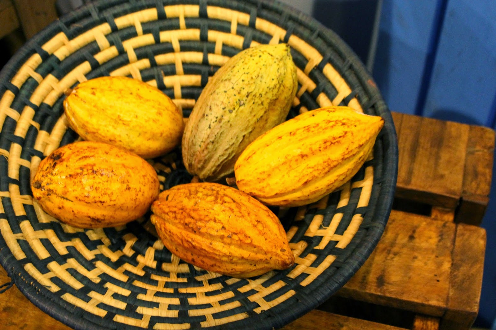 |
| Native cacao |
 |
| Highland Coffee |
Who said you can't grow food if you are living in a condo or without a land? there many ways and possibilities like this one, heirlomm seeds and herbs grown in a crates.


Let's preserve our heirloom and indigenous seeds, let's go organic, buy and go local, use locally available materials, plant and grow your own food if you can, support the local farmers in your area and encourage potluck, that way we do our little way of saving the environment.
To know more about Slow Food visit or contact:
ECHOSTORE
116 Tordesillas St., Salcedo Village, Makati City
+632 519.1216
E-mail: puj@echostore.ph
Website: www.echostore.ph
Wednesday, 13 August 2014
A Destiny We Cannot Understand
I have been reading about depression since Robin Williams died. The best article I read so far describes it as an energy, a tension that is stuck and appears to be immovable. The Buddhist teacher speaking about it says to merge with the depression, to become it, because it is so full of energy.
This seems like a big risk, doesn’t it? Instead of thinking of depression as something outside of us and afflicting us, we see it as an energy within us that has a lot of energy for us. When I think of this for myself, because I have dealt with depression on and off in the last six months, I feel a tremendous surge of energy that feels like anger well up inside me. Perhaps though it is not just anger, perhaps it is my power to heal, my power to create. Perhaps it is the divinity within me that has been bottled up and requires an outlet. A full expression of who I am.
I have been working with teachers and friends who are holding me accountable for being my most powerful self right now. In order to fulfill this destiny, I have to change, to be bigger and more fully realized. It feels so hard sometimes because I am not being supported to be my old small self anymore. Sometimes I feel abandoned because I am so addicted to that self that is needy and likes to be taken care of. That self is not really me. It is just an illusion of a kind of thinking that I let my mind do to get a kind of attention that feels comfortable.
The illusion of our thinking is so important in all this talk about Robin Williams. We don’t really know what his “mind chatter” was telling him. We don’t really know what his soul was calling for. We only know what we are told.
What if we accepted his death as what needed to happen for him?
When we can see through these eyes, we see that although sad, there is a kind of destiny here that we can not understand. And this mystery is what can open us to more expansive thinking.
To be supported to be bigger and to leave behind ways that no longer serve that goal, is the most powerful kind of support. It calls us into the realm of the mystery. This destiny for us into the unknown of so much beauty.
This seems like a big risk, doesn’t it? Instead of thinking of depression as something outside of us and afflicting us, we see it as an energy within us that has a lot of energy for us. When I think of this for myself, because I have dealt with depression on and off in the last six months, I feel a tremendous surge of energy that feels like anger well up inside me. Perhaps though it is not just anger, perhaps it is my power to heal, my power to create. Perhaps it is the divinity within me that has been bottled up and requires an outlet. A full expression of who I am.
I have been working with teachers and friends who are holding me accountable for being my most powerful self right now. In order to fulfill this destiny, I have to change, to be bigger and more fully realized. It feels so hard sometimes because I am not being supported to be my old small self anymore. Sometimes I feel abandoned because I am so addicted to that self that is needy and likes to be taken care of. That self is not really me. It is just an illusion of a kind of thinking that I let my mind do to get a kind of attention that feels comfortable.
The illusion of our thinking is so important in all this talk about Robin Williams. We don’t really know what his “mind chatter” was telling him. We don’t really know what his soul was calling for. We only know what we are told.
What if we accepted his death as what needed to happen for him?
When we can see through these eyes, we see that although sad, there is a kind of destiny here that we can not understand. And this mystery is what can open us to more expansive thinking.
To be supported to be bigger and to leave behind ways that no longer serve that goal, is the most powerful kind of support. It calls us into the realm of the mystery. This destiny for us into the unknown of so much beauty.
Saturday, 26 July 2014
Malunggay
 |
| Photo by: Lifestyle and Health. Malunggay (Moringa oleifera) – The Miracle Tree. April 13, 2014.[1] |
Benzolive, Drumstick Tree , Horse Radish Tree, Kelor, Marango, Mlonge, Mulangay, Saijhan and Sajna Moringa.[1]
Arunggai (Pang.), Balungai (P. Bis.), Dool (Bik.), Kamalongan (P. Bis.), Kalamungai (C. Bis.), Kalungai (Bik., Bis., Tag.), Kalunggay (Bik.), Kamalungai (Pamp., Tag.), Komkompilan (Ilk.), Molongai (Tag.), Malungay (Tag.), Malunggue (Pamp.), Malungit (Pamp., Bis.), Maroñgoi (Sbl.), Maruñgaai (Ilk., Ibn.), Drumstick tree (Engl.), Horse-radish tree (Engl.), La mu (Chin.), Ben oil tree (Engl.).[2]
Scientific names:
Moringa oleifera Linn., Moringa nux-ben Perr., Moringa pterygosperma Gaertn.., Guilandina moringa Linn.[2]
Descriptions:
Moringa oleifera, known popularly as “drumstick tree”, is an herbaceous plant grown for its nutritious leafy-greens, flower buds, and mineral-rich green pods. It is a well-recognized member in the “Moringaceae family” of trees, and thought to be originated in the sub-Himalayan ranges of Indian subcontinent. The plant possesses “horseradish-like root” and, hence, known to the western world as horseradish tree. Their young, tender seed pods are popular as “murnga” in Tamil, and “malunggay” in Philippine.[3]
This plant grows into a medium sized tree, 4 to 6 m tall. It can be kept to a useful size by regular pruning, and can be trained to grow as a hedge. The name drumstick comes from the distinctive long tapered seedpods that hang from the branches.[2]
Moringa oleifera Lam (Moringaceae) is a highly valued plant, distributed in many countries of the tropics and subtropics. It has an impressive range of medicinal uses with high nutritional value.[8]
Moringa has been identified as the vegetable with the highest nutritional value among many types of food species studied.
Availability: Drumstick trees are common in Fiji and Kiribati, but can be scarce in other Pacific islands and in northern Australia.[2]
Propagation methods: Plants can be produced from cuttings or seed; seed-derived plants are usually slower to establish but develop a strong root system. Cuttings of mature wood, 200 to 600mm long, planted with at least one-third of the cutting in the soil, are most suitable for propagation.[2]
How to grow: Drumstick trees are not difficult to grow. Once established, the tree is drought tolerant, can survive on shallow soil of poor fertility, will grow in full sun and is wind tolerant. The canopy of cuttinggrown plants can be pruned to increase wind tolerance. If growing conditions are poor, growth will be slower, and leaves smaller with a stronger flavour. For the first two years mulching is recommended, keeping the soil around the tree moist and free of grass and other weeds.
Threats: Pests and diseases are not usually a problem however root rot can occur if the tree is grown in waterlogged soils.
Harvesting: The leaves should be neatly picked, usually back to the third newest full leaf and ideally in the cooler hours of the day to prevent wilting.[2]
Post-harvest and storage: Full leaves (leaflets plus wiry stalks) should be washed carefully with water of drinking quality or clean seawater. If bundle wrapped in moist paper and kept in a cool location they should store for a day. Leaves can last for up to a week, if placed in an airtight container in a cool room or refrigerator. If the leaves dry they will drop their leaflets and lose their value as a food.
Environment:Originally from India, planted in frost free areas around the world. Naturalized in many areas. Grows best in sand soil, tolerates poor soil. It loves sun and heat and can be grown from seed.
Seed Pods: Immature seed pods are cooked and taken as food. It contains 0% cholesterol and very low fat but rich in dietery fibre,protein, energy, Vitamin A, Vitamin C, Thiamine, Riboflavin, Folate, Sodium, Pottasium, Calcium, Iron, Magnesium, Phosphorus, Selenium, and Zinc.
Flowers: Soup of flower is a good aphrodisiac. It can also be cooked and taken. Flowers contain Vitamin B, Vitamin B2, Vitamin B3, Vitamin C, Vitamin A in abundance.
Root: As traditional medicine, roots are used as gargle for painful gums and throat problems.
Roots are ground into paste and applied over glandular swellings.
Leaves: Leaves , flowers and pods is healthy for all. Include this miraculous food in your diet.
Plant parts utilized:
Roots[4], Seeds[4], Leaves[2],[4], Flowers[2], Young pods[2].
Picture of the plant’s important parts:
 |
| Photos by: [1]seed envy, [2]herbs India, [3]word press, [4]stuartxchange, [5]Dave's garden, [6]Christina Sarich. |
Active Constituents:
Malunggay contains the phytochemical niaziminin, which is found to have molecular components that can prevent the development of cancer cells and correlated with inhibitory ability against superoxide generation. The first naturally-occuring thiocarbamates, novel hypotensive agents niazinin A, niazinin B, niazimicin and niaziminin A and B were isolated from malunggay.[5]
Different parts of this plant contain a profile of important minerals, and are a good source of protein, vitamins, -carotene, amino acids and various phenolics. The Moringa plant provides a rich and rare combination of zeatin, quercetin, -sitosterol, caffeoylquinic acid and kaempferol. In addition to its compelling water purifying powers and high nutritional value, M. oleifera is very important for its medicinal value.[8]
Traditional use:Different parts of this plant contain a profile of important minerals, and are a good source of protein, vitamins, -carotene, amino acids and various phenolics. The Moringa plant provides a rich and rare combination of zeatin, quercetin, -sitosterol, caffeoylquinic acid and kaempferol. In addition to its compelling water purifying powers and high nutritional value, M. oleifera is very important for its medicinal value.[8]
Leaves contain[10]:
4 times more Vitamin A than Carrot
4 times more Calcium than Milk
2 times more Iron than Spinach
7 Times more Vitamin C than Oranges
3 times more Potassium than Banana
2 Times more protein than Eggs and Yogurt
For centuries, people in many countries have used Moringa leaves as traditional medicine for common ailments. Clinical studies have begun to suggest that at least some of these claims are valid. With such great medicinal value being suggested by traditional medicine, further clinical testing is very much needed. India: Traditionally used for anemia, anxiety, asthma, blackheads, blood impurities, bronchitis, catarrh, chest congestion, cholera, conjunctivitis, cough, diarrhea, eye & ear infections, fever, glandular swelling, headaches, abnormal blood pressure, hysteria, pain in joints, pimples, psoriasis, respiratory disorders, scurvy, semen deficiency, sore throat, sprain, tuberculosis.[1]
Malaysia: Traditionally used for intestinal worms.
Guatemala:Traditionally used for skin infections and sores.
Puerto Rico: Traditionally used for intestinal worms.
Philippines:Traditionally used for anemia, glandular swelling and Lactating.[1]
Traditional cultures in various parts of the world have long used Moringa in their herbal medicine repertoire for ailments ranging from gout to various inflammations and fevers.[9]
Therapeutic Activity[6]:Traditional cultures in various parts of the world have long used Moringa in their herbal medicine repertoire for ailments ranging from gout to various inflammations and fevers.[9]
Therapeutic Potential of M. oleifera in Chronic Hyperglycemia
Glucose homeostasis
Glucose is a major fuel for animal cells. It is supplied to the organism through dietary carbohydrates and, endogenously, through hepatic gluconeogenesis and glycogenolysis. Glucose absorption from the gastrointestinal tract (GIT) into blood is regulated by a variety of neuronal signals and enterohormones (incretins), as well as by meal composition and the intestinal flora. Glucose homeostasis reflects a balance between glucose supply and its utilization. Physiologically, this balance is determined by the level of circulating insulin and tissue responsiveness to it. Insulin is secreted by pancreatic islet β cells. It stimulates glucose uptake and utilization by tissues, especially by liver, skeletal muscle, and adipose tissue. It also suppresses gluconeogenesis in hepatocytes, while stimulating lipogenesis and inhibiting lipolysis in adipocytes (Gerich, 2000).
Hyperglycemia
An individual is diagnosed as diabetic when his blood glucose level is chronically ≥126 mg/dL after an overnight fast, and ≥200 mg/dL 2 h after an oral glucose load of 75 g (oral glucose tolerance test, OGTT; Alberti and Zimmet, 1998). Age, genetics, environment, and lifestyle influence the development of this pathology. The relative importance of these factors and their combinatorial effects are not yet fully understood. Two types of DM are commonly recognized: type 1 DM (T1DM) results from autoimmune destruction of pancreatic β cells and represents only 5% of all cases; type-2 DM (T2DM) is the most common form of the disease and the primary concern of this review.
In its early stages, T2DM is characterized by chronic hyperglycemia and hyperinsulinemia, due to loss of tissue sensitivity to insulin, and compensatory secretion of the hormone by islet β cells. Its progression involves a complex network of interacting cellular and physiological alterations leading to β cell failure. Glucotoxicity and lipotoxicity are the most commonly invoked mechanisms for this failure (Robertson et al., 2004).
Glucotoxicity arises from excessive uptake of glucose by islet β cells. The excess sugar drives glycation reactions and the mitochondrial electron transport chain, producing macromolecule-damaging reactive oxygen species (ROS), at levels beyond the antioxidation capacity of the cell. The ensuing oxidative stress impairs insulin synthesis and secretion, and initiates a cascade of cellular events that ultimately lead to apoptosis (Kaneto et al., 2007).
Lipotoxicity, on the other hand, results in part from the unresponsiveness of adipocytes to insulin, negating the ability of this hormone to stimulate uptake by these cells of non-esterified fatty acids (NEFA) that result from triglycerides (TG) lipolysis in circulation, and to inhibit lipolysis of endogenous TG to NEFA. Excess plasma NEFA impairs insulin secretion by β cells, stimulates gluconeogenesis by liver, and inhibits glucose disposal by skeletal muscle, further exacerbating hyperglycemia (Stumvoll et al., 2005). NEFA accumulation in the bloodstream is further aggravated by obesity, a condition characterized by an expanded adipose mass. Furthermore, adipose tissues, especially the visceral and deep subcutaneous ones, secrete pro-inflammatory cytokines such as interleukin 6 (IL-6) and tumor necrosis factor α (TNFα), which also contribute to tissue insensitivity to insulin.
Impaired TG storage into adipocytes facilitates the formation in the bloodstream of small, cholesterol ester-poor, TG-rich low-density lipoprotein (LDL) particles.
Hyperglycemia promotes glycation of these particles, a modification that extends their half-life in circulation. These particles are prone to oxidation and are potent initiators of atherogenesis and its vascular damages (discussed in more detail below). Diabetes-associated neuropathy, retinopathy, and nephropathy are some of the consequences of these damages (Dokken, 2008).
Underlying these complex physiological changes are molecular alterations in the relative levels of expression and post-translational modifications of a wide variety of gene products, including surface receptors, second messengers and transcriptional factors.
An individual is diagnosed as diabetic when his blood glucose level is chronically ≥126 mg/dL after an overnight fast, and ≥200 mg/dL 2 h after an oral glucose load of 75 g (oral glucose tolerance test, OGTT; Alberti and Zimmet, 1998). Age, genetics, environment, and lifestyle influence the development of this pathology. The relative importance of these factors and their combinatorial effects are not yet fully understood. Two types of DM are commonly recognized: type 1 DM (T1DM) results from autoimmune destruction of pancreatic β cells and represents only 5% of all cases; type-2 DM (T2DM) is the most common form of the disease and the primary concern of this review.
In its early stages, T2DM is characterized by chronic hyperglycemia and hyperinsulinemia, due to loss of tissue sensitivity to insulin, and compensatory secretion of the hormone by islet β cells. Its progression involves a complex network of interacting cellular and physiological alterations leading to β cell failure. Glucotoxicity and lipotoxicity are the most commonly invoked mechanisms for this failure (Robertson et al., 2004).
Glucotoxicity arises from excessive uptake of glucose by islet β cells. The excess sugar drives glycation reactions and the mitochondrial electron transport chain, producing macromolecule-damaging reactive oxygen species (ROS), at levels beyond the antioxidation capacity of the cell. The ensuing oxidative stress impairs insulin synthesis and secretion, and initiates a cascade of cellular events that ultimately lead to apoptosis (Kaneto et al., 2007).
Lipotoxicity, on the other hand, results in part from the unresponsiveness of adipocytes to insulin, negating the ability of this hormone to stimulate uptake by these cells of non-esterified fatty acids (NEFA) that result from triglycerides (TG) lipolysis in circulation, and to inhibit lipolysis of endogenous TG to NEFA. Excess plasma NEFA impairs insulin secretion by β cells, stimulates gluconeogenesis by liver, and inhibits glucose disposal by skeletal muscle, further exacerbating hyperglycemia (Stumvoll et al., 2005). NEFA accumulation in the bloodstream is further aggravated by obesity, a condition characterized by an expanded adipose mass. Furthermore, adipose tissues, especially the visceral and deep subcutaneous ones, secrete pro-inflammatory cytokines such as interleukin 6 (IL-6) and tumor necrosis factor α (TNFα), which also contribute to tissue insensitivity to insulin.
Impaired TG storage into adipocytes facilitates the formation in the bloodstream of small, cholesterol ester-poor, TG-rich low-density lipoprotein (LDL) particles.
Hyperglycemia promotes glycation of these particles, a modification that extends their half-life in circulation. These particles are prone to oxidation and are potent initiators of atherogenesis and its vascular damages (discussed in more detail below). Diabetes-associated neuropathy, retinopathy, and nephropathy are some of the consequences of these damages (Dokken, 2008).
Underlying these complex physiological changes are molecular alterations in the relative levels of expression and post-translational modifications of a wide variety of gene products, including surface receptors, second messengers and transcriptional factors.
Evidence of anti-hyperglycemic properties of M. oleifera
Moringa oleifera parts have been used in folk medicine for the treatment of diabetes (Dieye et al., 2008). Five studies aimed at verifying these properties using leaves were identified in the scientific literature: two were conducted in experimental animals (Ndong et al., 2007b; Jaiswal et al., 2009) and three in T2DM patients (William et al.,1993; Kumari, 2010; Ghiridhari et al., 2011). They are summarized in Table Table1.
 |
Table 1. Moringa oleifera experimental therapy for chronic hyperglycemia. |
In the study by Ndong et al. (2007b), Goto-Kakizaki (GK) Wistar rats were used as model of DM. GK rats spontaneously develop early glucose intolerance associated with impaired insulin secretion (Bisbis et al., 1993; Abdel-Halim et al.,1995). In an OGTT, overnight fasted Wistar controls or GK male rats were given 2 g/kg of body weight (kg-bw) glucose by oral gavage, without or with 200 mg/kg-bw of M. oleifera leaf powder. To determine blood glucose levels, vein blood was collected before gavage and at different times afterward up to 120 min. Areas under the curves (AUC) were derived from the time courses of these levels. In the absence of treatment, fasting plasma glucose levels (FPG) and their post-prandial levels (PPPG) at 120 min were greater (∼1.4× and 2.2×, respectively) in GK rats than in control rats. Treatment with M. oleifera leaf powder resulted in a lower glycemic response in GK and control rats. However, in GK rats, the treatment reduced AUC values by 23% (P < 0.05); it did not significantly affect these values in control rats. These observations suggested that M. oleifera treatment improves plasma glucose disposal only in the diabetic rats.
In the study by Jaiswal et al. (2009), prediabetes and diabetes were induced in Wistar rats by intraperitoneal (i.p.) injection of 55 mg/kg-bw of streptozotocin (STZ), a cytotoxic drug that selectively destroys islet β cells (Like and Rossini, 1976). Based on FPG (mg/dL), STZ-treated rats were classified as sub-diabetic (∼88 mg/dL), mildly diabetic (∼190 mg/dL), and severely diabetic (∼300 mg/dL). A M. oleifera aqueous extract was administered to overnight fasted animals by oral gavage, at 100, 200, or 300 mg/kg-bw. FPG was determined before treatment (baseline) and at various time points post-treatment. An OGTT was conducted 90 min after the last time point. In normal rats, M. oleifera treatment lowered FPG at all doses in time- and concentration-dependent manners. Six hours after administration, 200 mg/kg-bw of M. oleifera extracts lowered FPG by about 26% (P < 0.05), compared to baseline levels or the levels in untreated mice. In an OGTT, at the same dose, a 30% fall in PPPG was observed after 3 h, in normal, sub-diabetic, and mildly diabetic rats. A 21-day treatment of severely diabetic rats with the M. oleifera extract at a daily dose of 300 mg/kg-bw reduced FPG and PPPG by 69 and 51%, respectively, relative to untreated controls. In all the above experiments, the hypoglycemic effect of the plant extract was comparable to that of the anti-diabetic drug Glipizide administered at 2.5 mg/kg-bw.
Human studies
In a controlled study with untreated T2DM patients, William et al. (1993) examined how M. oleifera addition to a standardized meal, taken after an overnight fast, affected the 1- and 2-h PPPG, relative to the standard meal alone or a 75-g oral glucose load. M. oleifera was compared to bitter gourd (Momordica charantia) and curry leaves (Murraya koenigii). Compared to the glucose load, standard meals with or without vegetable supplements induced a significantly lower rise in PPPG (glycemic response) as derived from AUCs. However, when leaf-supplemented meals were compared to standard meals, only the M. oleifera leaf-supplemented meal elicited a lower response (−21%, P < 0.01). Plasma insulin AUCs did not differ significantly between the two meals, suggesting that the hypoglycemic effect of M. oleifera leaf supplementation was not due to increased insulin secretion.
Kumari (2010) examined the hypoglycemic effect of M. oleifera leaf dietary consumption over a 40-day period in T2DM patients, 30–60 years of age, not on anti-hyperglycemic medication. The experimental group included 46 subjects, 32 men, and 14 women; the control group of 9 subjects included 4 men and 5 women. Daily meals were comparable among these groups in terms of relative content of food types (e.g., cereals, green leafy vegetables, fruits, etc.) and nutrients (e.g., proteins, fat, fiber, minerals, etc.) as well as calories. The experimental group received a daily dose of 8 gM. oleifera leaf powder. FPG and PPPG at the end of the protocol (final) were compared to baseline levels. Final values did not differ much from baseline in the control group. They were significantly reduced in the experimental group (FPG: −28%, P < 0.01; PPPG: −26%, P < 0.05).
More recently, Ghiridhari et al. (2011) studied a group of 60 T2DM patients, age 40–58 years, BMI 20–25 kg/m2, on sulfonylurea medication and a standardized calorie-restricted diet. The patients were equally divided into an experimental and a control groups. Patients in the experimental group were prescribed two M. oleifera leaf tablets/day, one after breakfast, the other after dinner for 90 days. M. oleifera leaf powder constituted 98% (w/w) of the tablet content, but the average weight of tablets was not specified, making the total daily dose unclear. Blood glycated hemoglobin (HbA1c) was measured before and after the regimen. PPPG was determined before the regimen and every 30 days afterward. In the control group, HbA1c and PPPG progressed downwardly with time, but the change was not significant. In the experimental group, in contrast, relative to the baseline, HbA1c decreased by 0.4% point (from 7.8 ± 0.5 to 7.4 ± 0.6; P < 0.01). Compared to the starting levels (210 ± 49 mg/dL), PPPG in the experimental group progressively decreased with treatment duration, by 9% after 30 days, 17% after 60 days, and 29% after 90 days (P < 0.01), indicating that M. oleiferamedication can induce with time better glucose tolerance. However, it should be noted that treatment allocation to patients appear to have not been randomized as baseline values for the two parameters were higher in the experimental group than in the control group, 7.8 ± 0.5 vs. 7.4 ± 0.6% for HbA1c, 210 ± 49 vs. 179 ± 36 mg/dL for PPPG.
Therapeutic Potential of M. oleifera in Dyslipidemia
Lipid homeostasis
Lipids constitute a major class of hydrophobic constituents of the body. Their main forms are cholesterol, phospholipids (PL), and triglycerides (TG). Lipids are involved in a variety of biological processes, including membrane formation, intracellular and intercellular signaling, as well as energy storage and production. The body derives its lipids from de novo cellular biosynthesis and from nutrition. Cellular biosynthesis of lipids is regulated at the transcriptional level by sterol-regulated element-binding proteins (SREBPs) 1 and 2. SREBP-1 promotes the biosynthesis fatty acids and TG, SREBP-2 that of cholesterol (Horton, 2002).
Intestinal and plasma lipids are transported by lipoproteins particles. Apolipoproteins (Apo) constitute the protein components of these particles. Lipoproteins vary in density and, depending on their relative contents in TG, cholesterol, and PL, are identified as chylomicrons, very low-density lipoprotein (VLDL), LDL, intermediate density lipoprotein (IDL), and high-density lipoprotein (HDL). Lipids are transported by chylomicrons in the intestinal lymphatic system; and, in the bloodstream, by chylomicron remnants, VLDL, LDL, IDL, and HDL (Abeles et al., 1992; Havel and Kane,2001).
The liver plays a pivotal role in lipid metabolism. It extracts cholesterol from intestinal chylomicrons and excretes it back into the intestines with bile acids. It biosynthesizes TG and cholesterol and packages them as VLDL, that it secretes into the bloodstream. Through the LDL receptor (LDLR), it clears up plasma LDL as well as IDL from VLDL or HDL catabolism. HDL mediates the reverse transport of cholesterol from extra-hepatic tissues to the liver (Havel and Kane, 2001). Liver LDLR levels and its capacity to clear blood LDL are down-regulated by proprotein convertase subtilisin/kexin-type 9 (PCSK9), a plasma protein secreted by this organ (Horton et al., 2009).
Dyslipidemia
Dyslipidemia is a disorder characterized by alterations in the levels and composition of plasma lipids. According to Adult Treatment Panel III (2001), plasma levels ≥200 mg/dL for TC, ≥130 mg/dL for LDL-C, <40 mg/dL for HDL-C, and ≥150 mg/dL for TG are dyslipidemic. Dyslipidemia may result from inborn defects of lipoprotein production or metabolism; but in most cases, it is secondary to an unhealthy lifestyle (e.g., excessive cigarette smoking or alcohol consumption), other health disorders (e.g., obesity, diabetes, infection, obstructive liver disease), or medication (e.g., β blockers, steroids).
Besides hypertension, chronic dyslipidemia is a major cause of atherosclerosis, a vascular disease affecting blood circulation in the coronary, central, and peripheral arteries. The pathology is initiated by irritation of the arterial endothelium by high level of circulating LDL-C, which leads to overexpression of adhesion and chemoattraction molecules (e.g., vascular cell adhesion molecule-1, intercellular adhesion molecule, P and E selectins, monocyte chemoattractant protein-1) to injured sites, and the recruitment and capture of circulating monocytes to these sites. These immune cells penetrate into the sub endothelium and differentiate into tissue macrophages, which take up oxidized LDL (oxLDL) via scavenger receptors (e.g., CD36, scavenger receptor-A), becoming the lipid-laden foam cells characteristic of atheromatous plaques. In response to growth factors, resident vascular smooth muscle cells (VSMC) proliferate and form a fibrous cap overlying the plaques. The oxidative process that leads to oxLDL production also contributes to atherogenesis, as this modified lipoprotein and its by-products (oxysterols and oxPL) act as monocyte chemoattractants and VSMC mitogens. Clinical complications of this process include a narrowing of the arterial lumen, plaque rupture, and formation of circulating thrombi. These complications could lead to coronary artery disease (CAD), myocardial infarction, thrombo-embolic stroke, and peripheral artery disease (Steinberg, 2002; Libby et al., 2011).
Over the years, there has been a vigorous debate over the predictive value of plasma LDL-C level as a marker of CVD risk in humans. An emerging view is that the level of non-HDL lipoproteins, also captured in the TC/HDL-C or HDL-C/non-HDL-C ratio, may constitute a better marker, and its reduction a more cogent measure of the efficacy of anti-dyslipidemia therapies (Sniderman et al., 2010; Manickam et al., 2011).
Evidence of anti-dyslipidemic property of M. oleifera
Five studies were identified in the scientific literature: three were conducted with experimental animals (Ghasi et al., 2000; Chumark et al., 2008; Jain et al., 2010), two with human subjects (Kumari, 2010; Nambiar et al., 2010). They are summarized in Table 2.
Animal studies
Chumark et al. (2008) examined the therapeutic potential of M. oleiferaleaves on dyslipidemia induced in rabbits on a high-cholesterol (5%) diet (HCD) for 12 weeks. By the end of the regimen, relative to rabbits on a normal diet, HCD-fed rabbits experienced several-fold (×) increases in the plasma levels of total cholesterol (TC, 55×), HDL-C (17×), LDL-C (131×), and TG (4×). The diet also caused extensive plaque formation in carotid arteries. When these HCD rabbits were concomitantly fed a M. oleifera aqueous leaf extract, at the daily dose of 100 mg/kg-bw for the duration of the protocol, these increases were reduced: for TC and lipoprotein-cholesterol by about 50%, for TG by 75%, and for carotic plaque formation by 97%. This protective effect was comparable to that of the anti-cholesterol drug simvastatin, given p.o., at a daily dose of 5 mg/kg-bw. Similar results were also obtained with HCD rabbits fed an aqueous extract of M. oleifera fruits (Mehta et al., 2003).
The anti-dyslipidemic effects of M. oleifera leaves were also examined in rats fed a high-fat diet (HFD). In one study (Ghasi et al., 2000), Wistar rats were fed, for 30 days, a HFD containing 16% (w/w) fat, with or without an aqueous extract of M. oleifera leaves at a daily dose of 1 g/kg-bw. In untreated rats, the diet caused a 30% increase in plasma TC. In treated rats, the increase was reduced to 14%. In another study (Jain et al., 2010), albino rats were fed, for 30 days, a HFD containing 26% fat, with or without a methanolic extracts of M. oleifera leaves at daily doses of 150, 300, or 600 mg/kg-bw. In untreated rats, the diet increased plasma TC (2.4×), LDL-C (7.7×), VLDL-C (1.7×), and TG (1.6×). At the highest dose, M. oleifera treatment reduced these increases to 1.5×, 2.2×, 1.3×, and 1.3×, respectively (P < 0.01). Interestingly, serum HDL-C was unchanged by HFD diet alone; it was increased 2.4× in rats fed leaf extract-supplemented HFD, significantly reducing the TC/HDL-C ratio.
Human studies
Nambiar et al. (2010) examined the potential anti-dyslipidemic effect ofM. oleifera in 35 hyperlipidemic subjects (TC > 180 mg/dL or TG > 140 mg/dL), 26 men and 9 women. The control and experimental groups consisted of 18 subjects and 17 subjects, respectively. Anthropometric values (age, height, weight, body mass index, waist/hip ratio) within gender were similar between the two groups, as was their daily nutrient intake. The experimental group consumed a daily total of 4.6 g of dehydrated M. oleifera leaves, as four 550-mg tablets twice daily, for 50 days. Plasma lipid profiles were determined before and after the regimen. Compared to the control group, the experimental group experienced a 1.6% fall in plasma TC (P < 0.05) and a 6.3% increase of HDL-C, with non-significant trends toward lower LDL-C, VLDL-C, and TG. However, relative to baseline, final non-HDL-C and TC/HDL-C values decreased by 3.7 and 6.6%, respectively (P < 0.001), indicating that the treatment induced a lesser atherogenic lipid profile.
In the study of T2DM patients reported by Kumari (2010), the corrective effect of M. oleifera dietary leaves on dyslipidemia was also examined. Compared to the control group, the experimental group receiving 8 g of M. oleifera leaf powder daily for 40 days experienced a significant fall in the plasma levels of TC (−14%), LDL-C (−29%), VLDL-C (−15%), and TG (−14%; P < 0.05 to <0.01). HDL-C increased by 9% (non-significant), but the HDL-C/non-HDL-C ratio increased by 37% (P < 0.01).
Lipid homeostasis
Lipids constitute a major class of hydrophobic constituents of the body. Their main forms are cholesterol, phospholipids (PL), and triglycerides (TG). Lipids are involved in a variety of biological processes, including membrane formation, intracellular and intercellular signaling, as well as energy storage and production. The body derives its lipids from de novo cellular biosynthesis and from nutrition. Cellular biosynthesis of lipids is regulated at the transcriptional level by sterol-regulated element-binding proteins (SREBPs) 1 and 2. SREBP-1 promotes the biosynthesis fatty acids and TG, SREBP-2 that of cholesterol (Horton, 2002).
Intestinal and plasma lipids are transported by lipoproteins particles. Apolipoproteins (Apo) constitute the protein components of these particles. Lipoproteins vary in density and, depending on their relative contents in TG, cholesterol, and PL, are identified as chylomicrons, very low-density lipoprotein (VLDL), LDL, intermediate density lipoprotein (IDL), and high-density lipoprotein (HDL). Lipids are transported by chylomicrons in the intestinal lymphatic system; and, in the bloodstream, by chylomicron remnants, VLDL, LDL, IDL, and HDL (Abeles et al., 1992; Havel and Kane,2001).
The liver plays a pivotal role in lipid metabolism. It extracts cholesterol from intestinal chylomicrons and excretes it back into the intestines with bile acids. It biosynthesizes TG and cholesterol and packages them as VLDL, that it secretes into the bloodstream. Through the LDL receptor (LDLR), it clears up plasma LDL as well as IDL from VLDL or HDL catabolism. HDL mediates the reverse transport of cholesterol from extra-hepatic tissues to the liver (Havel and Kane, 2001). Liver LDLR levels and its capacity to clear blood LDL are down-regulated by proprotein convertase subtilisin/kexin-type 9 (PCSK9), a plasma protein secreted by this organ (Horton et al., 2009).
Dyslipidemia
Dyslipidemia is a disorder characterized by alterations in the levels and composition of plasma lipids. According to Adult Treatment Panel III (2001), plasma levels ≥200 mg/dL for TC, ≥130 mg/dL for LDL-C, <40 mg/dL for HDL-C, and ≥150 mg/dL for TG are dyslipidemic. Dyslipidemia may result from inborn defects of lipoprotein production or metabolism; but in most cases, it is secondary to an unhealthy lifestyle (e.g., excessive cigarette smoking or alcohol consumption), other health disorders (e.g., obesity, diabetes, infection, obstructive liver disease), or medication (e.g., β blockers, steroids).
Besides hypertension, chronic dyslipidemia is a major cause of atherosclerosis, a vascular disease affecting blood circulation in the coronary, central, and peripheral arteries. The pathology is initiated by irritation of the arterial endothelium by high level of circulating LDL-C, which leads to overexpression of adhesion and chemoattraction molecules (e.g., vascular cell adhesion molecule-1, intercellular adhesion molecule, P and E selectins, monocyte chemoattractant protein-1) to injured sites, and the recruitment and capture of circulating monocytes to these sites. These immune cells penetrate into the sub endothelium and differentiate into tissue macrophages, which take up oxidized LDL (oxLDL) via scavenger receptors (e.g., CD36, scavenger receptor-A), becoming the lipid-laden foam cells characteristic of atheromatous plaques. In response to growth factors, resident vascular smooth muscle cells (VSMC) proliferate and form a fibrous cap overlying the plaques. The oxidative process that leads to oxLDL production also contributes to atherogenesis, as this modified lipoprotein and its by-products (oxysterols and oxPL) act as monocyte chemoattractants and VSMC mitogens. Clinical complications of this process include a narrowing of the arterial lumen, plaque rupture, and formation of circulating thrombi. These complications could lead to coronary artery disease (CAD), myocardial infarction, thrombo-embolic stroke, and peripheral artery disease (Steinberg, 2002; Libby et al., 2011).
Over the years, there has been a vigorous debate over the predictive value of plasma LDL-C level as a marker of CVD risk in humans. An emerging view is that the level of non-HDL lipoproteins, also captured in the TC/HDL-C or HDL-C/non-HDL-C ratio, may constitute a better marker, and its reduction a more cogent measure of the efficacy of anti-dyslipidemia therapies (Sniderman et al., 2010; Manickam et al., 2011).
Evidence of anti-dyslipidemic property of M. oleifera
Five studies were identified in the scientific literature: three were conducted with experimental animals (Ghasi et al., 2000; Chumark et al., 2008; Jain et al., 2010), two with human subjects (Kumari, 2010; Nambiar et al., 2010). They are summarized in Table 2.
 |
Table 2. Moringa oleifera experimental therapy for chronic hyperlipidemia. |
Chumark et al. (2008) examined the therapeutic potential of M. oleiferaleaves on dyslipidemia induced in rabbits on a high-cholesterol (5%) diet (HCD) for 12 weeks. By the end of the regimen, relative to rabbits on a normal diet, HCD-fed rabbits experienced several-fold (×) increases in the plasma levels of total cholesterol (TC, 55×), HDL-C (17×), LDL-C (131×), and TG (4×). The diet also caused extensive plaque formation in carotid arteries. When these HCD rabbits were concomitantly fed a M. oleifera aqueous leaf extract, at the daily dose of 100 mg/kg-bw for the duration of the protocol, these increases were reduced: for TC and lipoprotein-cholesterol by about 50%, for TG by 75%, and for carotic plaque formation by 97%. This protective effect was comparable to that of the anti-cholesterol drug simvastatin, given p.o., at a daily dose of 5 mg/kg-bw. Similar results were also obtained with HCD rabbits fed an aqueous extract of M. oleifera fruits (Mehta et al., 2003).
The anti-dyslipidemic effects of M. oleifera leaves were also examined in rats fed a high-fat diet (HFD). In one study (Ghasi et al., 2000), Wistar rats were fed, for 30 days, a HFD containing 16% (w/w) fat, with or without an aqueous extract of M. oleifera leaves at a daily dose of 1 g/kg-bw. In untreated rats, the diet caused a 30% increase in plasma TC. In treated rats, the increase was reduced to 14%. In another study (Jain et al., 2010), albino rats were fed, for 30 days, a HFD containing 26% fat, with or without a methanolic extracts of M. oleifera leaves at daily doses of 150, 300, or 600 mg/kg-bw. In untreated rats, the diet increased plasma TC (2.4×), LDL-C (7.7×), VLDL-C (1.7×), and TG (1.6×). At the highest dose, M. oleifera treatment reduced these increases to 1.5×, 2.2×, 1.3×, and 1.3×, respectively (P < 0.01). Interestingly, serum HDL-C was unchanged by HFD diet alone; it was increased 2.4× in rats fed leaf extract-supplemented HFD, significantly reducing the TC/HDL-C ratio.
Human studies
Nambiar et al. (2010) examined the potential anti-dyslipidemic effect ofM. oleifera in 35 hyperlipidemic subjects (TC > 180 mg/dL or TG > 140 mg/dL), 26 men and 9 women. The control and experimental groups consisted of 18 subjects and 17 subjects, respectively. Anthropometric values (age, height, weight, body mass index, waist/hip ratio) within gender were similar between the two groups, as was their daily nutrient intake. The experimental group consumed a daily total of 4.6 g of dehydrated M. oleifera leaves, as four 550-mg tablets twice daily, for 50 days. Plasma lipid profiles were determined before and after the regimen. Compared to the control group, the experimental group experienced a 1.6% fall in plasma TC (P < 0.05) and a 6.3% increase of HDL-C, with non-significant trends toward lower LDL-C, VLDL-C, and TG. However, relative to baseline, final non-HDL-C and TC/HDL-C values decreased by 3.7 and 6.6%, respectively (P < 0.001), indicating that the treatment induced a lesser atherogenic lipid profile.
In the study of T2DM patients reported by Kumari (2010), the corrective effect of M. oleifera dietary leaves on dyslipidemia was also examined. Compared to the control group, the experimental group receiving 8 g of M. oleifera leaf powder daily for 40 days experienced a significant fall in the plasma levels of TC (−14%), LDL-C (−29%), VLDL-C (−15%), and TG (−14%; P < 0.05 to <0.01). HDL-C increased by 9% (non-significant), but the HDL-C/non-HDL-C ratio increased by 37% (P < 0.01).
Pharmacology of M. oleifera Leaves
Broad-spectrum physiological properties
Because of the chemical complexity of the M. oleifera medicinal formulations used in the studies reviewed above, their apparent therapeutic effects could be due to the combined actions of various bioactive components found in the plant, including trace metal ions, vitamins, alkaloids, carotenoids, polyphenols, fats, carbohydrates, and proteins (Coppin, 2008; Amaglo et al., 2010). Some compounds may collectively affect broad aspects of physiology, such as nutriment absorption and processing, redox state, or immunity.
Anti-nutrient properties
Moringa oleifera leaves contain phytosterols such as β-sitosterol (Jain et al., 2010). These compounds can reduce intestinal uptake of dietary cholesterol (Lin et al., 2010). They could partly account for the decrease of plasma cholesterol and the increase of fecal cholesterol observed in rodents treated with M. oleifera leaves (Mehta et al., 2003; Jain et al., 2010). M. oleifera leaf powder also contain about 12% (w/w) fibers (Joshi and Mehta, 2010). Dietary fibers reduce gastric emptying (Bortolotti et al., 2008). They may partly explain the greater stomach content, the improved OGTT response in treated GK diabetic rats (Ndong et al., 2007b), as well as the progressive improvement of PPPG levels in treated T2DM patients (Ghiridhari et al., 2011).
Antioxidant properties
The viability and functionality of a cell partly depends on a favorable redox state, i.e., on its ability to prevent excessive oxidation of its macromolecules, including DNA, proteins, and lipids (Ryter et al., 2007; Limon-Pacheco and Gonsebatt, 2009). ROS and free radicals are the major mediators of the oxidative process. Cellular inability to reduce ROS leads to oxidative stress. All cells are variably capable of endogenous self-protection against this stress through the actions of enzymes such as catalase, superoxide dismutase, and glutathione peroxidase, as well as through reducing molecules such as glutathione. Nutritional antioxidants such as vitamins A, C, and E provide additional protection from the stress (Limon-Pacheco and Gonsebatt, 2009).
Oxidative stress is widely accepted as a major contributing factor in the pathogenesis of CVD and diabetes (Dhalla et al., 2000; Kaneto et al., 2007; Rodrigo et al., 2011). A recurring explanation for the therapeutic actions of M. oleifera medication is the relatively high antioxidant activity of its leaves, flowers, and seeds (Chumark et al., 2008; Sreelatha and Padma, 2009; Verma et al., 2009; Atawodi et al., 2010). Among the major classes of phytochemicals found in the plant, flavonoids appear to carry most of this activity.
Anti-inflammatory properties
Inflammation with its wide array of cytokines secreted by immune cells is an integral part of the pathophysiology of obesity, hypertension, atherosclerosis, and diabetes (Rana et al., 2007). Extracts from M. oleifera leaves have been shown to modulate humoral and cellular immunity in rats and mice (Gupta et al.,2010; Sudha et al., 2010). They have exhibited strong anti-inflammatory properties in rodent models of chemically induced inflammation of the paw (Sulaiman et al., 2008; Mahajan and Mehta, 2009). These properties have been more extensively studied with fruit and seed extracts (Cheenpracha et al., 2010; Mahajan and Mehta, 2010; Muangnoi et al., 2011). They may also contribute to the observed anti-atherogenic and anti-diabetic effects of M. oleifera therapy.
Bioactive phytochemicals
An informative historical account of research in the phytochemistry of M. oleifera prior to 1995 can be found in Saleem’s doctoral thesis available on line (Saleem, 1995). Since then, the research has been expanded and refined, not only on the chemical structures of plant molecules, but also on their nutritional and medicinal properties. Of major medicinal interest are three structural classes of phytochemicals: glucosinolates, flavonoids, and phenolic acids (Saleem, 1995; Bennett et al., 2003; Lako et al., 2007; Manguro and Lemmen, 2007; Coppin, 2008; Amaglo et al., 2010; Kasolo et al., 2010). Their content in M. oleifera leaves varies somewhat with the geographic and climatic conditions under which the plant was grown, as well as with the processing methods for the collected leaves (Bennett et al., 2003; Coppin, 2008; Mukunzi et al., 2011).
Glucosinolates are characterized by β-thioglucoside N-hydroxysulfate motif (Figure (Figure2A).2A). In M. oleifera leaves, most phytochemicals of this class carry a benzyl-glycoside group linked to the single carbon of the motif. The most abundant of them is 4-O-(α-l-rhamnopyranosyl-oxy)-benzylglucosinolate, otherwise known as glucomoringin (Amaglo et al., 2010). Enzymatic hydrolysis of the glucosinolate motif of members of this class leads to the formation of corresponding isothiocyanates, thiocyanates, or nitriles. Several of these by-products have been shown to possess antihypertensive properties (Faizi et al., 1992, 1994, 1998).
Flavonoids and phenolic acids are collectively referred to phenolic compounds. The structural skeleton of flavonoids is made of two aromatic rings joined by a three-carbon link; that of the sub-class of flavonols is 3-hydroxy-2-phenylchromen-4-one (Figure (Figure2B).2B). Quercetin and kaempferol, in their as 3′-O-glycoside forms, are the predominant flavonols in M. oleifera leaves. The sugar moieties include, among others, rhamnoglycosyl (rutinosides), glucosyl (glucosides), 6′ malonyglucosyl, and 2′-galloylrutinoside groups (Bennett et al., 2003; Manguro and Lemmen, 2007; Amaglo et al., 2010). Biologically, flavonoids are best known for their antioxidant properties, but their metabolic pathways of activity remain to be fully elucidated (Rice-Evans, 2001). Phenolic acids have benzoic acid and cinnamic acid as backbones, with one or several hydroxyl groups (Figure (Figure2C).2C). Chlorogenic acid, which is an ester of dihydrocinnamic acid (caffeic acid) and quinic acid, is a major phenolic acid in M. oleifera leaves (Bennett et al., 2003; Amaglo et al., 2010).
Four of the best-characterized phytochemicals for their therapeutic efficacy in hyperglycemia, dyslipidemia, or related physiological conditions are shown in Figure Figure3.
Quercetin
Broad-spectrum physiological properties
Because of the chemical complexity of the M. oleifera medicinal formulations used in the studies reviewed above, their apparent therapeutic effects could be due to the combined actions of various bioactive components found in the plant, including trace metal ions, vitamins, alkaloids, carotenoids, polyphenols, fats, carbohydrates, and proteins (Coppin, 2008; Amaglo et al., 2010). Some compounds may collectively affect broad aspects of physiology, such as nutriment absorption and processing, redox state, or immunity.
Anti-nutrient properties
Moringa oleifera leaves contain phytosterols such as β-sitosterol (Jain et al., 2010). These compounds can reduce intestinal uptake of dietary cholesterol (Lin et al., 2010). They could partly account for the decrease of plasma cholesterol and the increase of fecal cholesterol observed in rodents treated with M. oleifera leaves (Mehta et al., 2003; Jain et al., 2010). M. oleifera leaf powder also contain about 12% (w/w) fibers (Joshi and Mehta, 2010). Dietary fibers reduce gastric emptying (Bortolotti et al., 2008). They may partly explain the greater stomach content, the improved OGTT response in treated GK diabetic rats (Ndong et al., 2007b), as well as the progressive improvement of PPPG levels in treated T2DM patients (Ghiridhari et al., 2011).
Antioxidant properties
The viability and functionality of a cell partly depends on a favorable redox state, i.e., on its ability to prevent excessive oxidation of its macromolecules, including DNA, proteins, and lipids (Ryter et al., 2007; Limon-Pacheco and Gonsebatt, 2009). ROS and free radicals are the major mediators of the oxidative process. Cellular inability to reduce ROS leads to oxidative stress. All cells are variably capable of endogenous self-protection against this stress through the actions of enzymes such as catalase, superoxide dismutase, and glutathione peroxidase, as well as through reducing molecules such as glutathione. Nutritional antioxidants such as vitamins A, C, and E provide additional protection from the stress (Limon-Pacheco and Gonsebatt, 2009).
Oxidative stress is widely accepted as a major contributing factor in the pathogenesis of CVD and diabetes (Dhalla et al., 2000; Kaneto et al., 2007; Rodrigo et al., 2011). A recurring explanation for the therapeutic actions of M. oleifera medication is the relatively high antioxidant activity of its leaves, flowers, and seeds (Chumark et al., 2008; Sreelatha and Padma, 2009; Verma et al., 2009; Atawodi et al., 2010). Among the major classes of phytochemicals found in the plant, flavonoids appear to carry most of this activity.
Anti-inflammatory properties
Inflammation with its wide array of cytokines secreted by immune cells is an integral part of the pathophysiology of obesity, hypertension, atherosclerosis, and diabetes (Rana et al., 2007). Extracts from M. oleifera leaves have been shown to modulate humoral and cellular immunity in rats and mice (Gupta et al.,2010; Sudha et al., 2010). They have exhibited strong anti-inflammatory properties in rodent models of chemically induced inflammation of the paw (Sulaiman et al., 2008; Mahajan and Mehta, 2009). These properties have been more extensively studied with fruit and seed extracts (Cheenpracha et al., 2010; Mahajan and Mehta, 2010; Muangnoi et al., 2011). They may also contribute to the observed anti-atherogenic and anti-diabetic effects of M. oleifera therapy.
Bioactive phytochemicals
An informative historical account of research in the phytochemistry of M. oleifera prior to 1995 can be found in Saleem’s doctoral thesis available on line (Saleem, 1995). Since then, the research has been expanded and refined, not only on the chemical structures of plant molecules, but also on their nutritional and medicinal properties. Of major medicinal interest are three structural classes of phytochemicals: glucosinolates, flavonoids, and phenolic acids (Saleem, 1995; Bennett et al., 2003; Lako et al., 2007; Manguro and Lemmen, 2007; Coppin, 2008; Amaglo et al., 2010; Kasolo et al., 2010). Their content in M. oleifera leaves varies somewhat with the geographic and climatic conditions under which the plant was grown, as well as with the processing methods for the collected leaves (Bennett et al., 2003; Coppin, 2008; Mukunzi et al., 2011).
Glucosinolates are characterized by β-thioglucoside N-hydroxysulfate motif (Figure (Figure2A).2A). In M. oleifera leaves, most phytochemicals of this class carry a benzyl-glycoside group linked to the single carbon of the motif. The most abundant of them is 4-O-(α-l-rhamnopyranosyl-oxy)-benzylglucosinolate, otherwise known as glucomoringin (Amaglo et al., 2010). Enzymatic hydrolysis of the glucosinolate motif of members of this class leads to the formation of corresponding isothiocyanates, thiocyanates, or nitriles. Several of these by-products have been shown to possess antihypertensive properties (Faizi et al., 1992, 1994, 1998).
 |
| Figure 2. Structural motifs and backbones of major phytochemicals found M. oleifera leaves. |
Four of the best-characterized phytochemicals for their therapeutic efficacy in hyperglycemia, dyslipidemia, or related physiological conditions are shown in Figure Figure3.
 |
| Figure 3. Some bioactive phytochemicals found M. oleifera leaves. |
The flavonol quercetin is found at concentrations as high as 100 mg/100 g of dried M. oleifera leaves (Lako et al., 2007), predominantly as quercetin-3-O-β-d-glucoside also known as isoquercitrin or isotrifolin (Bennett et al., 2003; Atawodi et al.,2010; Figure Figure3A).3A). Quercetin is a potent antioxidant (Zhang et al., 2011) with multiple therapeutic properties (Bischoff, 2008). It can reduce hyperlipidemia and atherosclerosis in HCD or HFD rabbits (Juzwiak et al., 2005; Kamada et al., 2005). It has shown anti-dyslipidemic, hypotensive, and anti-diabetic effects in the obese Zucker rat model of metabolic syndrome (Rivera et al., 2008). It can protect insulin-producing pancreatic β cells from STZ-induced oxidative stress and apoptosis in rats (Coskun et al.,2005). Its hypotensive effect has been confirmed in a randomized, double-blind placebo-controlled, human study (Edwards et al., 2007).
Chlorogenic acid
Chlorogenic acid (Figure (Figure3B)3B) can beneficially affect glucose metabolism. It has been shown to inhibit glucose-6-phosphate translocase in rat liver, reducing hepatic gluconeogenesis and glycogenolysis (Hemmerle et al., 1997; Karthikesan et al., 2010a). It was found to lower PPBG in obese Zucker rats (Rodriguez de Sotillo and Hadley, 2002). In OGTT experiments performed on rats or humans, it reduced the glycemic response in both species (van Dijk et al., 2009; Tunnicliffe et al.,2011); in rodents, it also reduced the glucose AUC (Tunnicliffe et al., 2011). Its anti-dyslipidemic properties are more evident as its dietary supplementation has been shown to significantly reduce plasma TC and TG in obese Zucker rats or HFD mice (Rodriguez de Sotillo and Hadley, 2002; Cho et al., 2010) and to reverse STZ-induced dyslipidemia in diabetic rats (Karthikesan et al., 2010b).
Moringinine
The alkaloid moringinine was initially purified from M. oleifera root bark (Ghosh et al., 1935) and later chemically identified as benzylamine (Chakravarti, 1955; Figure Figure3C).3C). It is also present in leaves. This substance was suspected to mediate the hypoglycemic effect of the plant. An early study showed that Wistar rats provided with drinking water containing 2.9 g/L of benzylamine for 7 weeks exhibited a reduced hyperglycemic response in an intraperitoneal glucose tolerance test (IPGTT), suggesting improved glucose tolerance (Bour et al., 2005). More recently, the effect was further explored using HFD-fed, insulin-resistant C57BL/6 mice taking an estimated daily dose 386 mg/kg-bw in drinking water for 17 weeks. Compared to untreated controls, these mice gained less weight, had reduced FPG and plasma TC, and were more glucose tolerant (Iffiu-Soltesz et al., 2010).
Niaziminin
Niaziminin (Figure (Figure3D)3D) is a mustard oil glycoside initially isolated (along with other glycosides such as niazinin and niazimicin) from ethanolic extracts of M. oleifera leaves, based on their hypotensive properties on Wistar rats. At 1 mg and 3 mg/kg-bw, these compounds caused a 16–22 and a 40–65% fall of mean arterial blood pressure (MABP), respectively (Faizi et al., 1992). Other active isothiocyanate glycosides and thiocarbamates were isolated from the plant using the same bioassay (Faizi et al., 1994, 1998; Saleem, 1995).
Aurantiamide acetate
This compound was isolated from M. oleifera roots and structurally identified as N-benzoylphenylalanyl phenylalinol acetate (Figure (Figure3E).3E). At 25 μM, this unusual dipeptide derivative inhibited by nearly 90% the secretion TNFα and IL-2 from lipopolysaccharide-stimulated peripheral blood lymphocytes in culture. It had no effect on IL-6 secretion (Sashidhara et al., 2009). This inhibitory activity may contribute to the anti-inflammatory properties of the plant.
Chlorogenic acid
Chlorogenic acid (Figure (Figure3B)3B) can beneficially affect glucose metabolism. It has been shown to inhibit glucose-6-phosphate translocase in rat liver, reducing hepatic gluconeogenesis and glycogenolysis (Hemmerle et al., 1997; Karthikesan et al., 2010a). It was found to lower PPBG in obese Zucker rats (Rodriguez de Sotillo and Hadley, 2002). In OGTT experiments performed on rats or humans, it reduced the glycemic response in both species (van Dijk et al., 2009; Tunnicliffe et al.,2011); in rodents, it also reduced the glucose AUC (Tunnicliffe et al., 2011). Its anti-dyslipidemic properties are more evident as its dietary supplementation has been shown to significantly reduce plasma TC and TG in obese Zucker rats or HFD mice (Rodriguez de Sotillo and Hadley, 2002; Cho et al., 2010) and to reverse STZ-induced dyslipidemia in diabetic rats (Karthikesan et al., 2010b).
Moringinine
The alkaloid moringinine was initially purified from M. oleifera root bark (Ghosh et al., 1935) and later chemically identified as benzylamine (Chakravarti, 1955; Figure Figure3C).3C). It is also present in leaves. This substance was suspected to mediate the hypoglycemic effect of the plant. An early study showed that Wistar rats provided with drinking water containing 2.9 g/L of benzylamine for 7 weeks exhibited a reduced hyperglycemic response in an intraperitoneal glucose tolerance test (IPGTT), suggesting improved glucose tolerance (Bour et al., 2005). More recently, the effect was further explored using HFD-fed, insulin-resistant C57BL/6 mice taking an estimated daily dose 386 mg/kg-bw in drinking water for 17 weeks. Compared to untreated controls, these mice gained less weight, had reduced FPG and plasma TC, and were more glucose tolerant (Iffiu-Soltesz et al., 2010).
Niaziminin
Niaziminin (Figure (Figure3D)3D) is a mustard oil glycoside initially isolated (along with other glycosides such as niazinin and niazimicin) from ethanolic extracts of M. oleifera leaves, based on their hypotensive properties on Wistar rats. At 1 mg and 3 mg/kg-bw, these compounds caused a 16–22 and a 40–65% fall of mean arterial blood pressure (MABP), respectively (Faizi et al., 1992). Other active isothiocyanate glycosides and thiocarbamates were isolated from the plant using the same bioassay (Faizi et al., 1994, 1998; Saleem, 1995).
Aurantiamide acetate
This compound was isolated from M. oleifera roots and structurally identified as N-benzoylphenylalanyl phenylalinol acetate (Figure (Figure3E).3E). At 25 μM, this unusual dipeptide derivative inhibited by nearly 90% the secretion TNFα and IL-2 from lipopolysaccharide-stimulated peripheral blood lymphocytes in culture. It had no effect on IL-6 secretion (Sashidhara et al., 2009). This inhibitory activity may contribute to the anti-inflammatory properties of the plant.
Toxicity[6]:
Hydroxyurea (HDU) is an antineoplastic agent that is commonly used in the treatment of Sickle cell disease (SCD).However, the therapeutic value of HDU is limited by its organotoxicity including testicular toxity. It has been shown that free radicals are involved in HDU-induced toxicity. The Application of natural phenolic compounds in the prevention of many pathologic diseases has been reported. Herein, the ability of polyphenolic-rich Moringa oleifera Leaf Extract (MOLE) to protect rat testis against HDU-induced histomorphometric, spermatogenic, and oxidative status impairments were investigated. Three experimental groups of Sprague-Dawley rats were used; MOLE- alone group that received orally MOLE 50 mg/kg body weight (b.w) daily for 90 consecutive days. HDU-alone group that had 25 mg HDU/kg b.w/day/orally for 90 consecutive days. MOLE plus HDU-group that were Moringa oleifera world’s most useful trees is also widely grown in the tropical regions.treated orally for 90 consecutive days with both 25 mg HDU/kg b.w/day and MOLE 50 mg/kg b.w/day. There was also a corresponding control group which had distilled water 2.5 ml/kg b.w/day/orally for 90 consecutive days. Our results demonstrated that co-treatment with MOLE protected the testis against the morphologic, spermatogenic and oxidative status changes induced by HDU. Industrial relevance: Hydroxyurea is commonly used in management of sickle cell disease. The use of this drug is however limited by its organotoxicity including testicular damage. The present study therefore explores the ability of extract of Moringa oleifera. Leaves to prevent testicular damage during HDU therapy with a view of providing base line information on its possible use as an adjunct in future treatment regimes.
REFERENCES:
[1] Dolcas Biotech LLC. (2006-2008). Nutritional Values of Moringa Leaves. Moringa: fresh leaf vs. dried leaf.
http://www.edlagman.com/moringa/moringa-fresh-leaf-vs-dried-leaf.pdf
[2] StuartXchange. August (2011). Malungay. Philippine Medicinal Plants.
[3] Umesh Rudrappa. (2009-14). Moringa Nutrition Facts. Power Your Diet: Your Guide to Healthier Nutrition.http://www.nutrition-and-you.com/moringa.html
[4] Luke Coutinho. (2013) Our Herbs. Herbs India: Pure Nutrition, Natural Treasures.
http://herbsindia.co.in/moringa_oleifera.html
[5]Mr. Adolf Victoria Ortega. (2010). food supplement: malunggay. Interchemex laboratories, inc.
http://www.interchemex.com/foods/malunggay.html
[6] Shamsuddeen Rufai, M. M. Hanafi, [...], and Jannatul Ferdous. May 21 (2013). Genetic Dissection of New Genotypes of Drumstick Tree (Moringa oleifera Lam.) Using Random Amplified Polymorphic DNA Marker. BioMed Research International.
http://www.ncbi.nlm.nih.gov/pmc/articles/PMC3687767/#sec1title
[5]Mr. Adolf Victoria Ortega. (2010). food supplement: malunggay. Interchemex laboratories, inc.
http://www.interchemex.com/foods/malunggay.html
[6] Shamsuddeen Rufai, M. M. Hanafi, [...], and Jannatul Ferdous. May 21 (2013). Genetic Dissection of New Genotypes of Drumstick Tree (Moringa oleifera Lam.) Using Random Amplified Polymorphic DNA Marker. BioMed Research International.
http://www.ncbi.nlm.nih.gov/pmc/articles/PMC3687767/#sec1title
[7] L C Saalu, A A Osinubi, A A Akinbami,O E Yama. (2011). Moringa oleifera Lamarck (drumstick) Leaf Extract Modulates the Evidences of Hydroxyurea -Induced Testicular Derangement. International Journal of Applied Research in Natural Products.
http://www.oalib.com/paper/2589163
[8] Green Deane. (2007 - 2014). Moringa, More Than You Can Handle. Eat The Weeds (and other things too).
http://www.eattheweeds.com/moringa-oleifera-monster-almost-2/
[8] Green Deane. (2007 - 2014). Moringa, More Than You Can Handle. Eat The Weeds (and other things too).
http://www.eattheweeds.com/moringa-oleifera-monster-almost-2/
[9] Fozia Farooq, Meenu Rai, Avinash Tiwari, Abdul Arif Khan and Shaila Farooq. (no date). Moringa oleifera: A plant with multiple medicinal uses and benefits. Moringa Medicinal Benefits.
http://miracletrees.org/moringa_medicine.html
[10] (no authur). (no date). Medicinal Uses or Health Benefits of Moringa Oleifera – Drumstick. Siddham.
http://siddham.in/medicinal-uses-or-health-benefits-of-moringa-oleifera-drumstick
Compiled by: Coriento, K. A. R.
http://miracletrees.org/moringa_medicine.html
[10] (no authur). (no date). Medicinal Uses or Health Benefits of Moringa Oleifera – Drumstick. Siddham.
http://siddham.in/medicinal-uses-or-health-benefits-of-moringa-oleifera-drumstick
Luyang Dilaw
 Luyang Dilaw
English Name: Turmeric
Common Name: Luyang Dilaw, Kalawag, Dilaw, Lampuyang, lawag[1]
Scientific Name: Curcuma longa [1]
Family Name: Zingiberidaceae [1]
Intoduction
Luyang dila is a perennial plant with roots or tubers oblong, palmate, and deep orange inside; root-leaves about 2 feet long, lanceolate, long, petioled, tapering at each end, smooth, of a uniform green; petioles sheathing spike, erect, central, oblong, green; flowers dull yellow, three or five together surrounded by bracteolae. It is propagated by cuttings from the root, which when dry is in curved cylindrical or oblong tubers 2 or 3 inches in length, and an inch in diameter, pointed or tapering at the end. [2] It is grown commercially in India and has been since before the written record. Unlike ginger, tumeric rhizome is brightly coloured, yellow as a matter of fact. The colour it produces is deep orange as compared to the canary yellow produced by saffron. It grew in India and was another import item for the Israelites. It was used by all the ancient cultures as both a food colouring and as a medicine. [3]
Part Used:
 |
| RHIZOME (root) |
Active Constituents
Turmeric constituents include the three curcuminoids: Curcumin (diferuloylmethane; the primary constituent and the one responsible for its vibrant yellow colour), demethoxycurcumin and bisdemethoxycurcumin, as well as volatile oils (tumerone, atlantone and zingiberone), sugars, proteins and resins. [4]
Traditional Use:
Many South Asian countries use it as an antiseptic for cuts, burns, and bruises, and as an antibacterial agent. In Pakistan, it is used as an anti-inflammatory agent, and as a remedy for gastrointestinal discomfort associated with irritable bowel syndrome and other digestive disorders. In Pakistan and Afghanistan, turmeric is used to cleanse wounds and stimulate their recovery by applying it on a piece of burnt cloth that is placed over a wound. Indians use turmeric, in addition to its Ayurvedic applications, to purify blood and remedy skin conditions. Turmeric paste is used by women in some parts of India to remove superfluous hair. Turmeric paste is applied to the skin of the bride and groom before marriage in some parts of India, Bangladesh, and Pakistan, where it is believed to make the skin glow and keep harmful bacteria away from the body. Turmeric is currently used in the formulation of several sunscreens. Several multinational companies are involved in making face creams based on turmeric. [5]
Another traditional use of turmeric is as a food colorant and dye for cloth – in both cases a cheaper alternative to saffron. It was and is used in religious ceremonies and offerings – often representing life, purity, and prosperity. [2]
Pharmacologic Activity
Internal
Antioxidant
Studies have shown that C. longa peel wastes possess antioxidant properties which could probably work by counteracting and or quenching of reactive oxygen species [6]
Alzheimer's Disease
Turmeric possesses multiple medicinal uses including treatment for AD. Curcuminoids, a mixture of curcumin, demethoxycurcumin, and bisdemethoxycurcumin, are vital constituents of turmeric. It is generally believed that curcumin is the most important constituent of the curcuminoid mixture that contributes to the pharmacological profile of parent curcuminoid mixture or turmeric. A careful literature study reveals that the other two constituents of the curcuminoid mixture also contribute significantly to the effectiveness of curcuminoids in AD Turmeric possesses multiple medicinal uses including treatment for AD. Curcuminoids, a mixture of curcumin, demethoxycurcumin, and bisdemethoxycurcumin, are vital constituents of turmeric. It is generally believed that curcumin is the most important constituent of the curcuminoid mixture that contributes to the pharmacological profile of parent curcuminoid mixture or turmeric. A careful literature study reveals that the other two constituents of the curcuminoid mixture also contribute significantly to the effectiveness of curcuminoids in AD. [7]
Colon Cancer
A recent study detailed the modulating effect of curcumin on apoptosis in tumors. Curcumin was administered to the test animals at 0.2% and 0.6% level in the diet late in the premalignant stage, during the promotion/progression stage of colon carcinogenesis in male rats. 0.2% curcumin significantly inhibited colon tumorigenesis in treated rats as compared to untreated controls. The inhibition of adenocarcinomas of the colon was found to be dose-dependent. The authors confirmed that the chemopreventive activity of curcumin is observed when it is administered prior to, during and after carcinogen treatment as well as late in the premalignant stage of colon carcinogenesis. [8]
Diabetes
Curcumin was given in different doses to SD rats after 4 weeks of diabetic GI complication induction. At the end of 4 weeks, significant GI dysfunction characterized by weight loss, delayed gastric emptying and intestinal transit associated with reduction in antioxidant enzyme levels and increased lipid peroxidation was observed. [9]
Weight loss
Curcumin was given in different doses to SD rats after 4 weeks of diabetic GI complication induction. At the end of 4 weeks, significant GI dysfunction characterized by weight loss, delayed gastric emptying and intestinal transit associated with reduction in antioxidant enzyme levels and increased lipid peroxidation was observed. [10]
Arthritis
Due to its anti-inflammatory and pain-relieving properties, it is not surprising that Turmeric is now being used as an effective natural remedy for Arthritis pain. Therefore, taking 500mg to 1000mg Turmeric capsules three times per day may provide significant relief from osteoarthritis pain. [10]
Asthma
Since turmeric is an anti-inflammatory, it can help reduce the inflammation associated with asthma. Add 1 teaspoon of turmeric powder to a glass of warm milk, and drink this mixture as an effective asthma home remedy. [10]
Cardiovascular disease
Age-related cardiovascular decline in postmenopausal women is characterized, in part, by increased left ventricular afterload, an indication of vascular dysfunction and hypertension. An 8 week pilot study randomized 45 postmenopausal women to one of four interventions: placebo, 150mg curcumin, exercise training plus placebo or exercise training plus curcumin. [11]
Gastrointestinal and Respiratory disorder
The crude extract of turmeric (Cl.Cr), relaxed the spontaneous and K+ (80 mM)-induced contractions in isolated rabbit jejunum as well as shifted the CaCl2 concentration-response curves. In rabbit tracheal preparation, Cl.Cr inhibited carbachol and K+-induced contractions. Activity directed fractionation revealed that the vasodilator and vasoconstrictor activities are widely distributed in the plant with no clear separation into the polar or non-polar fractions. When used for comparison, both curcumin and verapamil caused similar inhibitory effects in all smooth muscle preparations with relatively more effect against K+-induced contractions and that both were devoid of any vasoconstrictor effect and curcumin had no effect on atria. These data suggest that the inhibitory effects of Cl.Cr are mediated primarily through calcium channel blockade, though additional mechanism cannot be ruled out and this study forms the basis for the traditional use of turmeric in hyperactive states of the gut and airways. Furthermore, curcumin, the main active principle, does not share all effects of turmeric. [12]
Testicular Damage
The study demonstrated protective effects of low concentrations (1–50 μM) of curcumin on mouse sperm motility in vitro and on DEHP-induced damage of seminiferous tubules in testes and its ability to diminish the decrease in sperm motility in vivo. In contrast, curcumin used in high concentration (100 μM) decreased sperm motility and viability in vitro. The effects of curcumin were dependent on its concentration. In male germ cells in vivo the protective effect was seen despite the low bioavailability of curcumin. In contrast, high, unattainable in the organism, concentration of curcumin had a cytotoxic effect on male reproductive cells in vitro. Curcumin also had a protective effect against the harmful impact of DEHP on the male reproductive system. [13]
External
Dye
Precautions: Turmeric should be limited in women trying to get pregnant and should be avoided entirely hen pregnant. It should also be avoided by people with congestive heart failure. [14]
ToxicityIt has very low toxicity, too. As the global scenario is now changing towards the use of non-toxic plant products having traditional medicinal use, development of modern drugs from turmeric should be emphasized for the control of various diseases. Further evaluation needs to be carried out on turmeric in order to explore the concealed areas and their practical clinical applications, which can be used for the welfare of mankind. [15]
References
- http://www.stuartxchange.com/Dilaw.html
- Alter, Dean : Turmeric : http://www.herballegacy.com/Alter_History.html
- (2010) Doctor Schar : http://doctorschar.com/archives/tumeric-curcuma-longa/ya
- Yadav D, Yadav SK, Khar RK, Mujeeb M, Akhtar M. (2013): Turmeric (Curcuma longa L.): A promising spice for phytochemical and pharmacological activities. :http://www.greenpharmacy.info/article.asp?issn=0973-8258;year=2013;volume=7;issue=2;spage=85;epage=89;aulast=Yadav
- Sahdeo Prasad and Bharat B. Aggarwal (2011): Turmeric, the Golden Spice: Herbal Medicine: Biomolecular and Clinical Aspects. 2nd edition. :http://www.ncbi.nlm.nih.gov/books/NBK92752/
- M. Chethankumar, N. Anand, N.S. Gangadhara (11-2010) “Isolation and characterization of an antioxidant protein from turmeric (Curcuma longa L.) peel waste: A new biological source”, in Journal of Pharmacy Research. LA: Association of Pharmaceutical Innorvators. : http://warponline.org/uploads/contents/168-content-3-Preliminary-Assessment-of-In-vitro-Anticoagulant-Activity-vs.-Heparin-1,000I.U.-and-Cytotoxicity-of-Selected-Philippine-Medicinal-Plants.pdf
- Touqeer Ahmed and Anard- Hassan Gilani (07-19-2013) : Therapeutic Potential of Turmeric in Alzheimer's Disease: Curcumin or Curcuminoids?http://onlinelibrary.wiley.com/doi/10.1002/ptr.5030/abstract
- Sabinsa Corporation : (2009) :Curcumin C3 Complex http://curcuminoids.com/Pharmacological.htm
- Nitin Indarchandji Kochar, Kshitija Gonge, Anil V. Chandewar, C. D. Khadse : (2014) :Curcumin ameliorates gastrointestinal dysfunction and oxidative damage in diabetic rats: Internationa Journal o Pharmacological Research: http://ijpr.ssjournals.com/index.php/journal/article/view/86
- Apo Celestina de Alex (2013) : Health Benefits of Turmeric (Luyang Dilaw) : http://parakleto.com/index.php?do=/blog/623/health-benefits-of-turmeric-luyang-dilaw/
- Sugawara J, Akazawa N, Miyaki A, Choi Y, Tanabe Y, Imai T, Maeda S. (06-2012): Effect of endurance exercise training and curcumin intake on central arterial hemodynamics in postmenopausal women: pilot study. Am J Hypertens.: Turmeric (Curcma longa) : http://www.drlise.net/attachments/tumeric.pdf
- Anwarul Hassan Gilani, Abdul Jabbar Shah, Muhammad Naeel Ghayur, Kashif Majeed(2005) :Pharmacological basis for the use of turmeric in gastrointestinal and respiratory disorders : Life Sciences: http://www.sciencedirect.com/science/article/pii/S0024320505000950
- Kataryza Glombik, Agnieszka basta-Kaim, Marta Sikora- Polaczek, Marta Kubera, Gabriela Starowicz, Jozefa Styma, (2014) :Curcumin influences semen quality parameters and reverses the di(2-ethylhexyl)phthalate (DEHP)-induced testicular damage in mice : Pharmacological Reports : http://www.sciencedirect.com/science/article/pii/S1734114014001662
- Specialty Herb Store : http://www.specialtyherbstore.com/Turmeric_Powder_Curcuma_longa_p/bhturm.htm
- Hamid Nasri, Najmeh Shahinfard, Mortaza Rafieian, Samira Rafieian, Maryam Shirzad, Mahmoud Rafieian : (2014) : TURMERIC: A SPICE WITH MULTIFUNCTIONAL MEDICINAL PROPERTIES : Journal of Herbmed Pharmacology :http://herbmedpharmacol.com/index.php/herb/article/view/45
Compiled By: Casuyon, Mel Rose S.
Subscribe to:
Posts (Atom)